aura · biophotons · faint light
Living tissue gives off a faint light. The old name for that field is aura.
You came here because you wanted the glow around a body to be real.
You already had a name for that glow. People have called it an aura for a long time, and they still do. Labs later counted a faint light that living tissue already makes. You can keep the aura name and still hear what those labs counted.
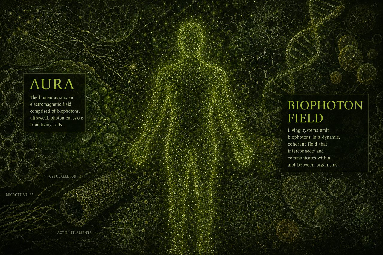
What is an aura?
An aura is the old name for a field around a living body. People who work with energy already talk about that field. They say you can feel it near the skin, see a color, or sense a person before they speak.
You want that field to be real. The lab work here is a count of faint light from living tissue.
If you already use the old maps of the body, open the chakras page and the meridians page next.
What is a biophoton?
A photon is a tiny packet of light. Ultraweak means that packet is too faint for your eyes. A biophoton is that faint light from a living body. Labs also call the same count ultraweak photon emission, or UPE.
Living tissue gives off a few to several hundred of those packets each second on each square centimeter of skin. A square centimeter is about the size of a fingernail. Labs see that light between 200 and 800 nanometers. A nanometer is how long a light wave is, a billionth of a meter, so those numbers name colors from near ultraviolet, the light just past what you can see, through the colors your eyes already know.
Mould and colleagues reviewed this in Frontiers in Physiology in 2024. They treat the faint count as a real thing living tissue already does. You can read that review on the journal site.
Alexander Gurwitsch ran onion-root work in 1923. He pointed one root at another through quartz, a clear stone that lets that kind of light through, and he called the effect mitogenetic radiation. That phrase means a kind of light that seemed to change how cells divide. Fritz-Albert Popp coined the word biophoton in 1984. He said DNA might be a source, and he guessed the light might stay in step with itself. That guess is still a guess.
Can you see it?
You cannot see this light with your eyes, because the count is too faint for the cells in the back of your eye. People can see it with dark-room cameras. A photomultiplier is a dark-room camera that counts single bits of light. A CCD is another dark-room camera that builds a picture from those bits. People put a body in a dark room and let those cameras count.
The pictures here are drawings that help you hold the idea. Do not treat the boxes on the art as a count from a camera.
What did Kobayashi measure?
Masaki Kobayashi, Daisuke Kikuchi, and Hitoshi Okamura put five people in a dark room in 2009. They used a CCD, a dark-room camera that counts single bits of light, and watched the face and the torso. They saw more light on the face than on the torso. They counted about 3000 photons each second on each square centimeter of cheek at 16:00, about double the 10:00 count. Diurnal means the count changes through the day.
When they took a heat picture of the same five people and laid it next to the light picture, the warm places and the bright places did not match, so heat and glow were two different maps of the same body.
Van Wijk and Van Wijk counted 29 sites on the body in 2005. They found the lowest counts on the chest and the highest counts on the head and the hands. Cohen and Popp wrote in 2003 about a rhythm and a left-right pattern, which means the two sides of the body can move together through the day. Ives and colleagues reviewed ultraweak photon emission as a health check in 2014 and called the idea promising.

Is a Kirlian photo the same thing?
A Kirlian photo is a photo made with high voltage on a plate. Corona is the spark around that plate. Semyon Kirlian put an object on film, ran high voltage, and a halo showed up. People still sell that halo in shops as the field around a person.
Pehek, Kyler, and Faust showed in Science in 1976 that moisture on the skin drove most of that picture. When your skin is wet, the spark changes. When you press harder, the spark changes. You can want a picture of the field around a person and still keep a phone photo made with high voltage on a plate apart from the faint light labs count in a dark room with no plate at all.
Is DNA a laser?
Popp said DNA might be a source, and he guessed the light might stay in step the way a laser does. A laser is a tight beam of light that stays in step with itself. Popp offered that as an idea. Clinics do not run your body as a laser.
Pietruszka and Marzec measured ultraweak photon emission from barley DNA in Scientific Reports in 2024. They suggest DNA is a major source of that faint light. Volodyaev and Beloussov wrote in 2015 that the coherent-field claim, the idea that the whole body runs as one tight light field, had not got experimental proof yet.
When your cells use oxygen, they can make extra light. People call that chemistry oxidative. They name the busy pieces ROS, which means reactive oxygen species, the pieces you get from burning oxygen. When you are stressed, those pieces can rise, and the count can rise with them. It does not turn your body into a handheld laser.
If you want the rise people feel in the chest, open the kundalini page. If you want the vacuum and the records, open the akashic page.
What do you do with this now?
You came here because you wanted the glow around a body to be real. That is a fair thing to want.
Living tissue already gives off a faint light. People used aura as the name for that field long before a dark-room camera could count it. Keep the name.
A shop photo made with high voltage on a plate is a spark. The labs counted something quieter. You do not need a dark-room camera to care about the field. Keep the aura name. The labs counted a faint light living tissue already makes.
Sources
Mould, R.R., Mackenzie, A.M., Kalampouka, I., Nunn, A.V.W., Thomas, E.L., Bell, J.D., & Botchway, S.W. “Ultra weak photon emission: a brief review.” Frontiers in Physiology 15:1348915 (14 Feb 2024). UPE as metabolic photons, a few to several hundred per second per square centimeter, about 200 to 800 nm. ROS as currently accepted source. Popp’s coherent-regulation vision still waits on a full experimental proof. doi.org/10.3389/fphys.2024.1348915
Gurwitsch, A. “Die Natur des spezifischen Erregers der Zellteilung.” Archiv fur mikroskopische Anatomie und Entwicklungsmechanik 100:11-40 (1923). Onion-root mitogenetic radiation through quartz. Cited via Mould 2024.
Popp, F.A., Nagl, W., Li, K.H., Scholz, W., Weingartner, O., & Wolf, R. “Biophoton emission. New evidence for coherence and DNA as source.” Cell Biophysics 6:33-52 (1984). Non-thermal UV-visible, DNA as candidate source. PMID 6204761. doi.org/10.1007/BF02788579
Kobayashi, M., Kikuchi, D., & Okamura, H. “Imaging of ultraweak spontaneous photon emission from human body displaying diurnal rhythm.” PLoS One 4(7):e6256 (2009). Five people. Face brighter than torso. Cheeks about 3000 photons per second per square centimeter at 16:00, about double 10:00. Thermograph did not match. PMID 19606225. doi.org/10.1371/journal.pone.0006256
Van Wijk, E.P.A., & Van Wijk, R. “Multi-site recording and spectral analysis of spontaneous photon emission from human body.” Forschende Komplementarmedizin und Klassische Naturheilkunde 12(2):96-106 (2005). 29 sites. Chest lowest. Head and hands highest. PMID 15947468. pubmed.ncbi.nlm.nih.gov/15947468
Cohen, S., & Popp, F.A. “Biophoton emission of human body.” Indian Journal of Experimental Biology 41:440-445 (2003). Rhythm and left-right. PMID 15244265.
Pietruszka, M., & Marzec, M. “Ultra-weak photon emission from DNA.” Scientific Reports 14:28915 (2024). Barley genomic DNA. Authors suggest DNA is a major UPE source. doi.org/10.1038/s41598-024-80469-0
Volodyaev, I., & Beloussov, L.V. “Revisiting the mitogenetic effect of ultra-weak photon emission.” Frontiers in Physiology 6:241 (2015). Coherent-field claim still waits on experimental proof. doi.org/10.3389/fphys.2015.00241
Pehek, J.O., Kyler, H.J., & Faust, D.L. “Image modulation in corona discharge photography.” Science 194(4262):263-270 (1976). Moisture as the main driver of Kirlian images. High-voltage corona on a plate. PMID 17836780. doi.org/10.1126/science.968480
Ives, J.A., van Wijk, E.P.A., Bat, N., et al. “Ultraweak photon emission as a non-invasive health assessment: a systematic review.” PLoS One 9(2):e87401 (2014). PMID 24586244. doi.org/10.1371/journal.pone.0087401